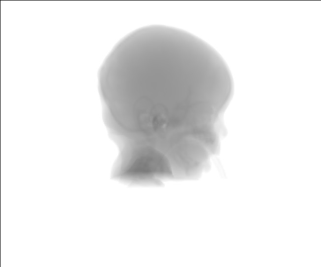
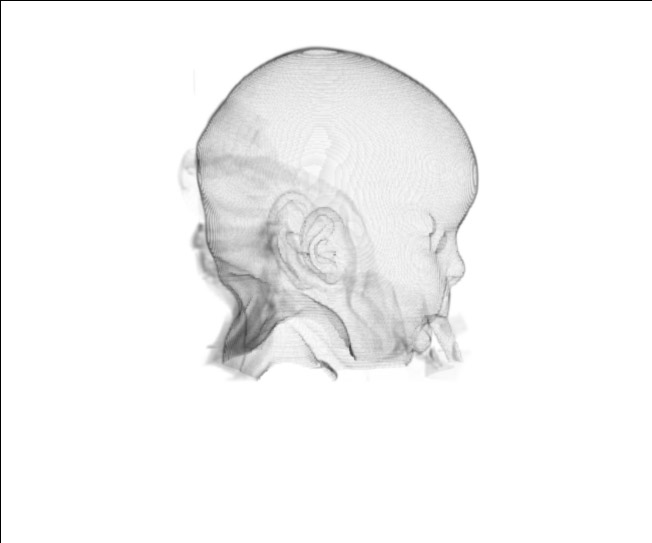
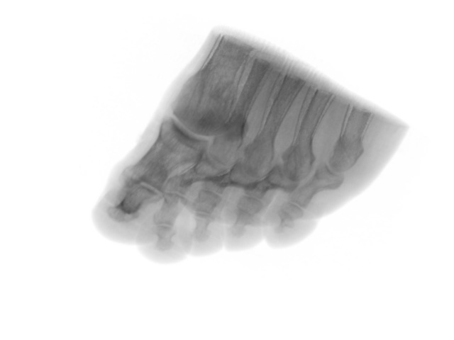
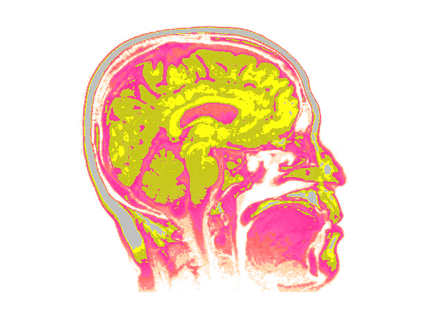
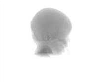
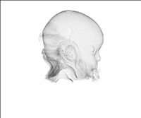
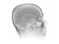
In Figure 1 we present CT- and MR-derived volumes. The data was taken from radiology.uiowa.edu and www.volren.org.
a) CT of the human skull;
b) CT of the human skull with filtration resulting in skin showing;
с) Sagittal MR image of the brain;
d) CT of the foot.
a)
b)
voxel size in mm - 0.35x0.35x2, resolution - 256x256x97

c)
d)
| voxel size in mm - 0.98x0.98x3 |
voxel size in mm - 1.00x1.00x1.00 |
| resolution - 256x256x19 |
resolution - 256x256x256 |
Figure 1
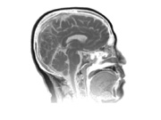
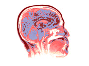

Figure 2. Vizualizing of slice. Color is a function of density.
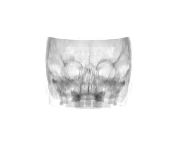
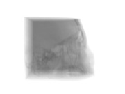
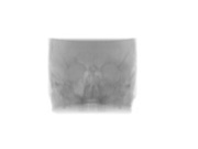
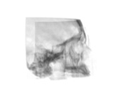
Figure 3 depicts CT images of the human skull in different planes using various filters to accent soft tissue.
a, b) CT of the skull;
с, d) CT of the skull: using filters to extract bones tissue.

a)
b)
c)
d)
Figure 3. Voxel size in mm - 0.7x0.7x2, resolution - 256x256x203
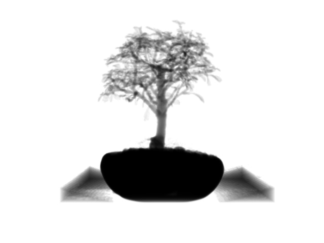
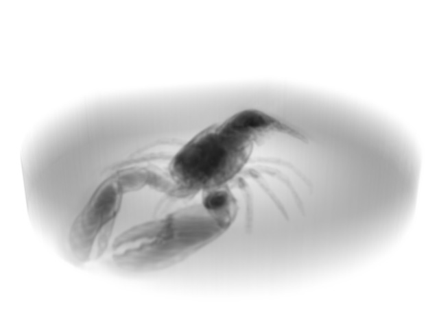
Figure 4 shows CT image of bonsai tree (left) and lobster (right).
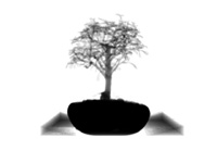

| voxel size in mm - 1.00x1.00x1.00 |
voxel size in mm - 1.00x1.00x1.40 |
| resolution - 256x256x256 |
resolution - 301x324x56 |
Figure 4
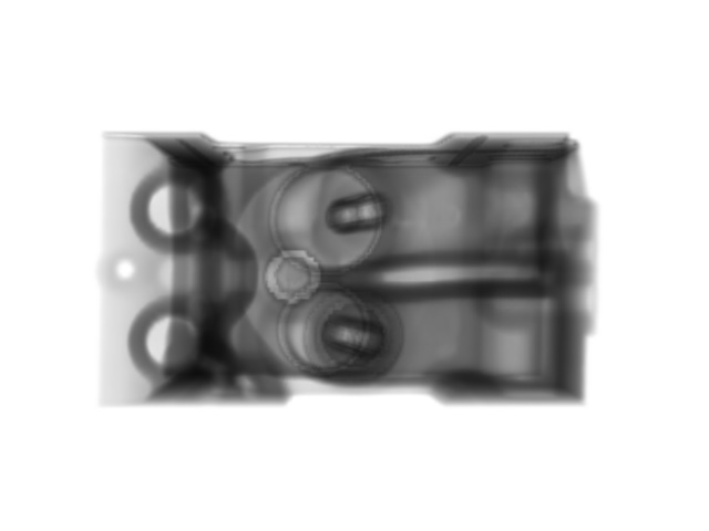
Intravision, or volume visualization, has applications in technical sciences as well. In Figure 5 we present CT images of cylinder block head in different planes.
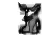
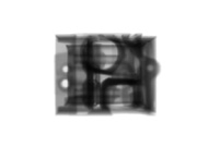

Figure 5. Voxel size in mm - 1.00x1.00x1.00, resolution - 256x256x256.
|